In the realm of eye health, accurately diagnosing color vision deficiencies, such as color blindness, has historically been a challenge. Before the advent of modern diagnostic tools, many individuals with color vision issues went undiagnosed. This was particularly problematic in fields where color discrimination is crucial, such as in textiles, fashion design, and various forms of visual arts. Recognizing colors correctly is not just about seeing the world more vividly; it’s essential for effective communication and quality assurance in color-critical professions.
The Breakthrough of the Ishihara Test
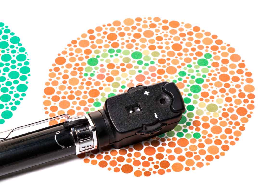
Enter the Ishihara test, a revolutionary approach to detecting red-green color deficiencies, developed by Dr. Shinobu Ishihara in 1917. This test transformed the way professionals could diagnose and understand color blindness. It uses a series of plates filled with dots of different colors and sizes to form numbers or shapes. These designs are visible to those with normal color vision but may be invisible or difficult to decipher for those with a red-green color deficiency.
How Does the Ishihara Test Work?
The Ishihara test consists of various plates, each designed to evaluate the viewer’s ability to perceive color differences.
- It is a color perception test that uses 38 plates in order to detect red-green color deficiencies. It is also called as ‘38 plates CVD test’. There are several types of plates that are given to the person under test.
- Each of the plates consists of a circle of dots that appear to be randomized in color and size.
- Some plates contain a number which is visible to the person with normal eyes and invisible or very difficult to the person with red-green color deficiencies.
- Other plates contain numbers that are visible to the person with deficient eyes and barely visible to those with normal eyes.
- The results extracted from plate observations define the person as normal or deficient. Normally, a severe deficiency can be detected after a few plates and a little deficiency may require all plates to be categorized accordingly.
Types of Plates
There are several types of plates that are given to the person under test. The test includes:
- Transformation Plates: These show different images or colors to viewers depending on their color vision status.
- Vanishing Plates: Only those with normal color vision can see the figures on these plates.
- Hidden Digit Plates: These are designed to be visible only to those with color vision deficiencies.
- Diagnostic Plates: These help determine the severity and type of color blindness, distinguishing between conditions like protanopia and deuteranopia.
The test is typically administered under controlled lighting conditions, with the plates presented at a specific distance and angle. The ability to correctly identify the numbers or shapes on the majority of the plates indicates normal color vision, while difficulty or inability to do so suggests a color vision deficiency.
Test Method and Environment
- The plates are designed to be assessed in a room that is lit adequately by daylight or artificial lamp close to daylight.
- Plates should be held at a distance of 75 cm from the subject and tilted so that the plane of the paper is at right angles to the line of vision.
- The plates 1–25 should be read within 3 seconds.
- If the person is unable to read the numerical, plates 26–38 are used and the winding lines between the two ‘X’ are traced with the brush.
- Each tracing should be completed within 10 seconds.
- An assessment of the readings of plates 1–21 determines the normality or color vision defect.
- If 17 or more plates are read normally, the color vision is regarded as being normal.
- If 13 plates (or less) are read correctly, the color vision is regarded as deficient.
- Testing with the first 24 plates gives a more accurate diagnosis of the severity of the color vision defect.
- Ishihara color plates nos. 4, 6, 10 and 16 read as numerical 29, 5, 2 and 16 respectively by a normal observer.
- Common plates include a circle of dots in shades of green and light blues with a figure differentiated in shades of brown, or a circle of dots in shades of red, orange and yellow with a figure in shades of green.
Explanation of The Plates
 Loading…
Result Analysis
As assessment of the reading of plates 1 to 21 determines the normality or defectiveness of color vision. If 17 or more plates are read normally, the color vision is regarded as normal. If only 13 or less than 13 plates are read normal, the color vision is regarded as deficient.
- However, in reference to plates 18, 19, 20, and 21, only those who read the numerals 5, 2, 45, and 73 and read them easier than those on plates 14, 10, 13 and 17 are recorded as abnormal.
- It is rare to find a person whose recording of normal answers is between 14-16 plates. An assessment of such a case requires the use of other color vision tests, including the anomalscope.
- In the assessment of color appreciation by the short method involving 6 plates only as described on page 4, a normal recording of all plates is proof of normal color vision.
- If there is a discrepancy in any of the recordings, the full series of plates should be used before diagnosing a red-green deficiency.
Why Is the Ishihara Test Important?
The Ishihara test is not just a medical curiosity; it’s a critical tool for identifying individuals who may face challenges in professions where color discrimination is essential. Early detection of color vision deficiencies can help individuals make informed career choices and seek out resources to manage their condition effectively.
Moreover, in our increasingly visual world, understanding and accommodating color vision deficiencies is important for creating accessible designs in everything from digital content to public spaces. The Ishihara test plays a vital role in raising awareness and understanding of color blindness.
Making the Test Accessible
Today, the Ishihara test is widely available, including online versions that offer a convenient way for individuals to screen for color vision deficiencies from the comfort of their homes. While these online tests can provide valuable insights, they are not a substitute for professional diagnosis and evaluation by an eye care specialist.
Conclusion
The Ishihara test remains a cornerstone in the field of optometry for diagnosing color blindness. Its simplicity, combined with its effectiveness, makes it an invaluable resource for ensuring that color vision deficiencies are recognized and properly addressed. Whether you’re a professional whose career depends on accurate color perception or someone curious about your color vision status, the Ishihara test offers a clear window into how you see the world.
Understanding and addressing color vision deficiencies is more than a medical concern; it’s about ensuring that everyone can experience the world in its full vibrancy and participate fully in work and daily activities that depend on accurate color perception.
You can test yourself online using the below link.
References
S. Ishihara, Tests for color-blindness (Handaya, Tokyo, Hongo Harukicho, 1917). Kindel, Eric. “Ishihara”. Eye Magazine. Retrieved 3 December 2013. Fluck, Daniel. “Color Blindness Tests”. Colblinder. Retrieved 3 December 2013. “Whonamedit – dictionary of medical eponyms” www.whonamedit.com. Retrieved 12 August 2015. www.dfisica.ubi.pt/~hgil/p.v.2/Ishihara/Ishihara.24.Plate.TEST.Book.pdf www.colorblindness.com/ishihara_cvd_test/ishihara_cvd_test.html